It used to be that if you received a diagnosis of glaucoma, chances were good that you would eventually lose vision to some degree. That began to change with the development of drugs that lower intraocular pressure, elevated levels of which can drive glaucoma. The medications were by no means a cure-all, but in people for whom the drugs worked it could buy time, slowing the deterioration of the visual field, or the extent of the area they could see.
Over the years, laser and surgical techniques evolved that could also slow disease progression. In a trabeculectomy, the surgeon creates an opening, or vent, for fluid to drain out of the eye, thus lowering eye pressure. In other approaches, a small tube shunt or stent is implanted in the eye to facilitate the drainage of fluid. Over the last decade, the advent of minimally invasive glaucoma surgery (MIGS) has meant minimal trauma and faster recovery times.
Even with these advances, however, there is a high rate of complications following glaucoma surgery: Shunts can drain too much fluid initially, jeopardizing vision. Fibrosis, or scar tissue, can occur around shunts, slowing or halting the drainage of fluid — especially problematic with the tiny devices used in MIGS. Shunts can also become clogged from naturally occurring proteins in the eye. Inflammation can cause the shifting of implants.
In an attempt to overcome such complications, surgeons have developed various workarounds. For example, when implanting a tube shunt the surgeon may place a suture inside the bore, or lumen, of the tube to prevent the sudden release of too much fluid. The suture degrades over time, at which point the body will (hopefully) have built up enough scar tissue around the site to slow the flow of fluid through the shunt. Still, up to 45 percent of glaucoma surgeries will fail within five years, leaving patients at risk of vision loss.
If Not Here, Then Where?
As a glaucoma specialist at Wilmer Eye Institute, Ian Pitha knew firsthand the limitations of glaucoma surgery. One day, as Pitha was leaving the OR and reflecting on a particularly challenging case, he stopped by the lab of Laura Ensign, Wilmer’s vice chair of research. “I remember thinking, with all the resources I have here, we really should be able to address this,” he recalls. Pitha described his frustration to Ensign, who suggested that he talk to Kunal Parikh, a biomedical engineering doctoral student at Johns Hopkins University who was working on novel medical device applications using materials with a unique set of properties.
As a chemical engineering major, Parikh had learned a technique known as electrospinning, a method of manufacturing nanofibers and microfibers. Early on, scientists created mats out of the fibers, which were used for everything from water filtration and purification to surgery to repair torn rotator cuffs. Today these nanofibers are being manufactured for a range of applications, including sutures, cardiac stents and vascular grafts.
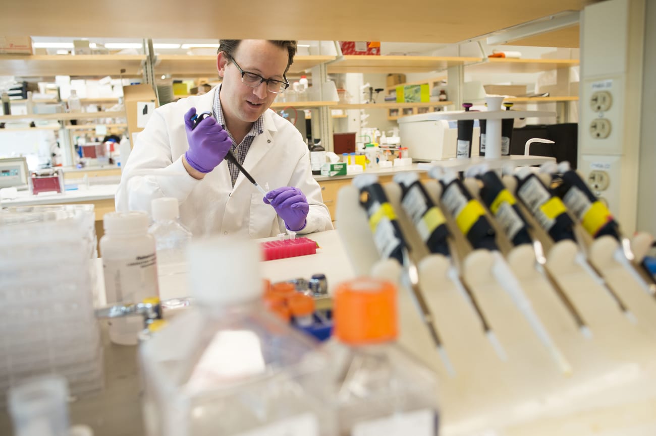
“A unique aspect of nanofibers is that they mimic the natural extracellular matrix that cells are around all the time,” says Parikh. “We're no longer just make mats, but now we're making all sorts of different embodiments that can have a big impact, primarily because of the way that cells interact and integrate with these types of nanofiber-based devices,” he says.
Working in the Center for Nanomedicine at Wilmer, Parikh and his colleagues had developed a new system for manufacturing a variety of nano-structured medical devices. It was here that Pitha met Parikh, who was leading a project to create drug-eluting sutures. Previous attempts to load drug directly into a suture had led to the sutures being too weak, but by twisting nanofibers into a multi-filament suture, Parikh and his team were able to load medication in the suture without the suture losing its mechanical properties.
Pitha was fascinated. He and Parikh began meeting regularly to discuss the current challenges of glaucoma surgery — challenges that might be amenable to solving with this technology.
Testing a Hypothesis
Pitha and Parikh worked together to define their goals. They wanted to see if porous stents made out of nanofibers would provide an advantage over the comparatively smooth materials that were currently utilized in glaucoma drainage implants. Would they, like the mats used to repair rotator cuffs, better integrate with the body’s own biology, thereby minimizing inflammation and the buildup of scar tissue? There was also data showing that nanofibers can prevent protein absorption, which often results in clogged stents. Would this bear out for glaucoma stents?
They decided to test their hypothesis, creating a nanofiber stent and implanting it in a rabbit eye. Parikh explains that many times, with any sort of implant, cells will deposit tissue around the stent in an attempt to encapsulate it and segregate it from the rest of the body. “They perceive it as foreign — a threat. That's what leads to that fibrotic response. What we found here was that we didn't see that encapsulation. Instead we saw cells actually migrate into the wall of the stents,” says Parikh.
The fact that the cells actually went directly into the stent demonstrated biocompatibility and validated the team’s hypothesis that the nanofibers provide a more natural environment for cells. Moreover, because the cells integrated directly within the wall of the stent, the stent didn't migrate.
Embracing the Spirit of Discovery
Energized by their findings, Pitha and Parikh wondered how they might further improve the outcomes of glaucoma surgery using nanotechnology. For example, what could they do to address the risk of plummeting eye pressure when a stent is first implanted? They went on to develop a second-generation stent with a degradable inner core that provides initial resistance to control fluid outflow, but then degrades as the body heals.
The pair published their findings in 2020. A second study, aimed at gaining a deeper understanding of the benefits of using nanofibers in comparison to smooth surfaces, is nearing completion. That study also looks at retention of the nanofiber structure over time.
Recently, Pitha and Parikh, along with engineer Steven Storck from the Johns Hopkins University Applied Physics Lab, received a Johns Hopkins Discovery Award that will allow them to further develop their technology for MIGS. “The people from the APL have a lot of expertise in modeling and fabrication, utilizing resources we don’t have,” says Pitha, who sees many additional possibilities for the use of nanomaterials in the treatment of glaucoma. For example, currently there are no commercially available stents that provide drug delivery, which could vastly improve the outcomes of glaucoma surgery.
Both Pitha and Parikh laud the Discovery Awards as being integral to their research. “The spirit of these Discovery Awards is enabling these multidisciplinary collaborative efforts, and I think this is a really great example of that, says Parikh.
But they also point to the collaborative environment at Wilmer itself. “It’s great that this was purely built off the clinical need and the resources that are available here. It was born from a clinical frustration and then a realization that if I’m not doing it in here, then I don’t know who’s going to do it anywhere else,” says Pitha.